
Essays and evidence on ageing, from LONGEVITY’s doctors and specialists.
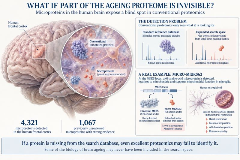
The ageing proteins we were not looking for
Proteomics can only identify what its search space allows. A human brain study highlights tiny, overlooked proteins and asks[…]

The most dangerous medical misinformation begins with the truth
A patient brought an advert that looked like a medical explanation. Following its claims back to the research revealed[…]
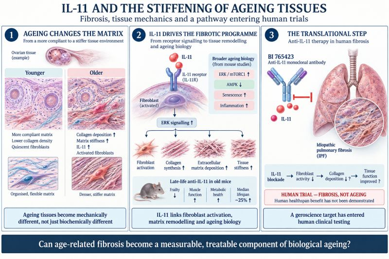
IL-11 and the stiffening of ageing tissues
Ageing changes the physical environment in which cells live. Research on IL-11 links tissue stiffness, fibrosis and loss of[…]
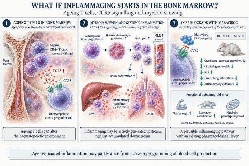
Does inflammaging start in the bone marrow?
Ageing T cells may influence the immune cells our bone marrow produces. A mouse study involving CCR5 blockade offers[…]
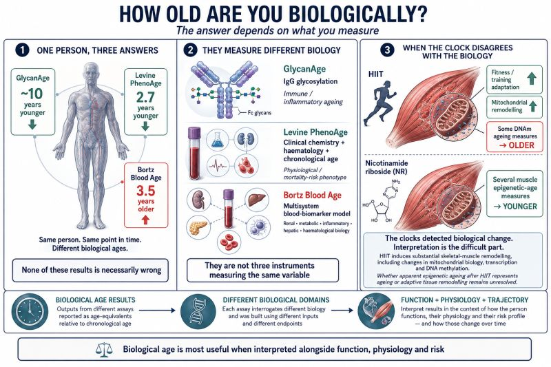
When biological age clocks disagree
Ten years younger by one test, older by another. Disagreement between biological age measures can be informative, provided we[…]
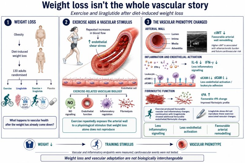
Weight loss is not the finished job
Getting lighter is useful. Becoming stronger, fitter and metabolically healthier is a larger ambition. Research combining exercise and liraglutide[…]

© 2026 Longevity Clinics Europe (UK) Limited